Você está usando um navegador desatualizado. Atualize seu navegador para uma experiência mais veloz e segura.

Carregando...

Atendimento via WhatsApp

Reconstructive Urology

Unique Approach, Multidisciplinary Team

At the Reconstructive Urology Center led by Prof. Tiago Rosito, we have built a unique approach to the reconstruction of the genital and urinary tract based on a multidisciplinary team. With the collaboration of urologists, plastic surgeons, colorectal surgery, gynecology, physical therapy, and nursing, we aim to maximize the functional and cosmetic outcomes of our reconstructive surgeries. As a pediatric and reconstructive urologist, Prof. Tiago Rosito performs surgeries aimed at improving quality of life, alleviating patient suffering, and often providing transformative benefits.

Areas of Expertise        Specialties    

Specialties in Reconstructive Urology

When a patient loses a significant amount of genital tissue due to trauma, burns, or infection, genital reconstruction surgery can help restore anatomy and minimize scarring. The genitals are traditionally difficult to reconstruct and often require a combination of muscle flaps, skin grafts, or even tissue expander balloons placed under the skin. Reconstruction of the urinary tract involves reconstructing the structures that store or transport urine-namely, the bladder, urethra, and ureter.

Bladder reconstruction usually involves increasing the size of the bladder with a portion of the small intestine or colon and then bringing the urine to the skin through a surgically constructed channel that can be continent (catheterizable channel/Mitrofanoff) or incontinent. When the bladder is completely removed, we can recreate a "new" bladder with small intestine (neobladder) in an orthotopic manner by reconnecting it to the native urethra or in a heterotopic manner through an abdominal conduit (ileal pouch).

Urethral reconstruction involves a combination of skin grafts, oral mucosa grafts, and skin and muscle flaps to increase the urethral diameter so that urination can be restored to normal.

Ureteral reconstruction typically involves filling the gap of the absent or diseased ureter by replacing the ureter with an elongated bladder, a bladder tube, or a segment of the small intestine or appendix to act as a chimney to transport urine to the bladder.

Laparoscopy and robotic surgery are suitable and minimally invasive options for ureteral and bladder reconstruction and help minimize hospitalization and overall recovery time. Robotic reconstruction is an evolving and expanding field, and at iPROURO, we are committed to using the most advanced surgical techniques. We foresee that most abdominal urinary reconstructions will become less invasive over time and with our growing experience.

Reconstructive urology is an evolving field; many of the procedures we commonly perform today did not exist ten years ago. We embrace innovation and collaboration. With a reflective and open mindset, we continually strive to improve and refine our functional and cosmetic outcomes.

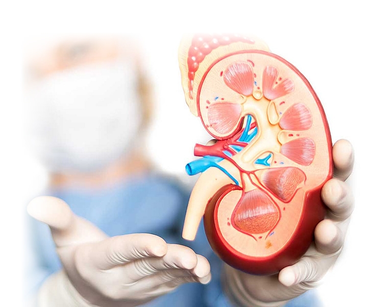
 

Congenital defects affect the urinary tract more commonly than any other organ system. These anomalies may affect only one portion, such as the urethra (hypospadias), or they may manifest as a constellation of congenital problems involving multiple organ systems. Common urological conditions that affect multiple organs are: spina bifida (myelomeningocele), exstrophy-epispadias complex, and cloacal malformations. These conditions are often accompanied by long-standing problems with bladder and/or bowel storage and emptying-which can severely affect a person's social life and overall quality of life.

Due to recent advances in neonatal, pediatric, and surgical care, many children with complex congenital diseases are able to survive into adulthood, compared to previous decades. In fact, more than 90% of patients with spina bifida can now live into adulthood. As the total number of children with complex urological conditions surviving into adulthood has grown dramatically, the transition of care for such patients from the pediatric urologist to the adult urologist is a pressing and urgent issue.

Most pediatric patients are able to find an adequate level of care in a pediatric hospital, but adults with the same conditions often lose follow-up and struggle to find quality care. Patients who do not transition to adult care are at a much higher risk of increased kidney damage, urinary tract infections, sepsis, and stones, as well as dramatically increased healthcare costs.
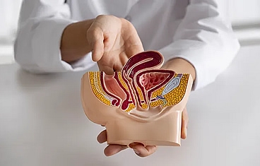
As children with congenital urological problems become adults, issues of sexuality, post-pubertal genital appearance and function, urinary and fecal control, fertility, and pregnancy become much more important to the patient and become important components of a full and independent life. Additionally, as they age, patients with congenital urogenital anomalies may develop the typical age-related urological problems, such as erectile dysfunction, prostate diseases, and even prostate, bladder, and kidney tumors.

Professor Tiago Rosito presents a contemporary profile of managing these patients, a complete and qualified urologist for comprehensive care from childhood to adulthood with the goal of maximizing quality of life and independence for adults born with congenital urological problems. Working at one of the country's leading medical and research facilities (Hospital de Clínicas de Porto Alegre and Hospital Moinhos de Vento), Professor Tiago Rosito is recognized as one of the most important national and international specialists in reconstructive urology.

Areas of Expertise in Reconstruction

Quality of Life Post Neoplasia

In our urological reconstruction group, we consider quality of life as important as the quantity of life.

The ProUro Cancer Survivorship Program is a unique and much-needed multidisciplinary program that addresses quality of life issues before, during, and after cancer therapy.

Complications of cancer therapy can be severe and include sexual dysfunction, urinary loss, and infertility. Often, in the effort to prolong life, these possible complications are not considered until therapy ends. With our multidisciplinary approach, we help patients make choices before starting cancer therapy that can help them avoid serious complications. When complications of therapy occur, we have the latest surgical techniques and support team available to resolve them.

Our team also specializes in penile and urethral preservation surgeries that can avoid emasculation in the treatment of genital cancer.

As a result of advances in early detection and treatment, the number of patients living well beyond a cancer diagnosis has greatly increased and will continue to grow in the foreseeable future. The American Cancer Society estimated that in 2012, there were 13.7 million cancer survivors in the US, and by 2022, the number of survivors will approach 18 million. Additionally, increased awareness of symptoms, better technology, and adherence to screening protocols have helped increase the number of cancer survivors. Quality of life choices become increasingly important as more survivors deal with the complications of cancer treatment.

Damage to the genitourinary tract often comes from the treatment of cancers of the pelvis and retroperitoneum (the back of the abdomen) - namely colorectal, gynecological, and urological cancers. Complications can include male stress incontinence (urine loss), rectourethral fistulas, urethral strictures, bladder neck contractures, genital mutilation and emasculation, ureteral strictures, urinary diversion, and sexual dysfunction - all potentially devastating and life-altering for the cancer survivor.

Our primary goal is to help our patients make choices that maximize quality of life before, during, and after cancer treatment.

Adult Hypospadias

Hypospadias is a congenital condition (present at birth) in which the penile urethra does not close properly during its development. Children born with hypospadias have a urethra that does not extend to the end of the head of the penis. Instead, the urethra ends short and on the body of the penis. Most hypospadias urethras have the opening on the underside of the penis and not at the tip. However, in some very severe cases, the urethra ends in the scrotum or behind it.

The urethra is the tube that carries urine from the bladder to the outside of the body. It also serves as the channel through which semen is ejaculated.

Most often, hypospadias is diagnosed shortly after birth. Your child will need surgery to correct the defect ideally around 1 year of age. Depending on the location of the urethral opening, the closer to the scrotum, the more difficult its reconstruction.

What kind of problems do adults born with hypospadias have?

Many patients with hypospadias reach adulthood and continue to have problems urinating or chronic urine infections. Common problems are urethral stenosis, urethral fistula (hole in the skin), hair in the urethra (from a previous reconstruction that used hairy skin), urethral stones, urinary tract infections, and fertility difficulties.
Patients who reach adulthood with complications from childhood surgical repairs often have complex problems that require additional complex repairs. These repairs usually require a combination of grafts from the inner lining of the cheek and skin flaps. Often, more than one surgery is needed to reconstruct adult patients with complex hypospadias, particularly because local or reliable skin is absent after unsuccessful childhood surgeries. It is essential to have an evaluation by a surgeon experienced in this pathology during all stages of male development.

Adult Spina Bifida, Myelomeningocele, and Bladder Exstrophy

Spina bifida is a birth defect involving incomplete closure of the spine and the membranes around the spinal cord. The most common location is the lower back. Common problems associated with spina bifida include bladder and/or bowel control problems, swelling of the brain ventricles (hydrocephalus), tethered spinal cord, latex allergy, and difficulty walking. To help manage all these complex problems, we have established a detailed holistic approach to care.
Bladder exstrophy (BE) is a congenital problem in which the bladder does not close. The open bladder is exposed on the abdominal wall. The pelvic bones are also abnormal and widely separated. The prostate and penis (epispadias) are also commonly abnormally formed. Bladder exstrophy is a rare birth defect and occurs in one in every 10,000 to 50,000 live births. Reconstruction usually involves closing the bladder and abdominal wall, repairing the flattened penis, and preventing urine leakage. At the time of repairing urine leakage, the bladder may be augmented with a piece of intestine if necessary.

Bladder Reconstruction for Cancer or Trauma

The bladder can be surgically reconstructed based on whether the entire bladder or part of the bladder is surgically removed and whether the urethra or bladder neck is scarred or cancerous. If the entire bladder is removed due to cancer, a new bladder can be made from the small intestine. This can happen at the same time as cancer surgery or at a later time. To build a bladder, 60 to 70 cm of the small intestine is opened and sewn into a spherical sac. The new bladder is then sewn to the urethra. While the neobladder is healing, urine is typically drained by 2 bladder catheters and 2 ureteral stents.

After 3 weeks, an X-ray of the bladder is performed to confirm that it is well healed. The catheters are then removed, and the patient is instructed on how to urinate. The new bladder is only a flexible urine pouch and therefore can only be emptied with the effort of the abdominal muscles. These small intestine pouches are often called "Hautmann" or "Studer" pouches. If part of the bladder is removed, it can be reconstructed by augmenting the bladder with the small intestine or the right colon. Sometimes the urethra or bladder neck is severely scarred or there is cancer in the urethra. In these cases, a catheterizable channel of the intestine can also be brought to the umbilicus or abdominal wall. This constructed channel is called an Indiana Pouch or Mitrofanoff procedure.

Congenital Penile Curvature

Congenital curvature of the penis during erection is commonly called "chordee." The penis usually curves downward. Patients who present with penile curvature throughout life do not experience pain during erection. However, erections with curvatures greater than 15 degrees may make penetration during sexual intercourse difficult, and some women complain of discomfort or pain due to the abnormal curvature.

Chordee repair requires a surgical approach where permanent sutures are placed to straighten the erect penis. The outpatient surgery usually takes one hour. The risks to the quality of the erection are almost nil. The success rate of chordee repair reaches 95%.

Chordee should not be confused with Peyronie's disease.

Genital Lymphedema

Lymphedema involves the accumulation of fluid in the skin, making it thick and leathery. Because of gravity, fluid buildup is usually worse in the lower parts of the body, such as the scrotum and legs. The amount of fluid that can accumulate in the genital skin can be considerable. In severe cases, genital lymphedema can be painful and debilitating and can make walking and movement difficult.

The diagnosis of genital lymphedema usually requires ruling out other possible causes for genital swelling, such as congestive heart failure or pelvic cancer. If no clear cause can be determined and the amount of discomfort is high, a surgical approach can achieve excellent functional and cosmetic results. Very thick and heavy genital skin can be surgically removed and grafted onto the skin of the penis and scrotum to unbury it. The results are usually excellent, and patients can achieve good functional outcomes that can improve walking and urination, as well as an acceptable cosmetic penis.

Urinary Diversions and Catheterizable Conduits

When the bladder is removed due to cancer or trauma, the urinary tract needs to be reconstructed to carry urine from the abdomen to the outside of the body. This is called urinary diversion.

After the bladder is removed or enlarged, the options for controlling urine are:

Ileal conduit - a piece of small intestine sewn to the skin where urine constantly drains, requiring an ostomy appliance "attached" to the skin to collect urine.

Orthotopic neobladder - a new bladder created from the small intestine, sewn to the urethra (where urine is expelled outside through the urethra).

Heterotopic neobladder - where a urine pouch is created outside the intestine, stores urine, and is regularly drained to the outside by periodically passing a catheter through the skin into the pouch. The most common surgically constructed pouch by our group is the Continent Ileal Conduit.

It is a continent, catheterizable urine pouch. The urine pouch is made of intestine - specifically the ileum.

The ileum is reconfigured into a U-shape, used to enlarge the bladder or to create a new reservoir. A flap of this ileal loop is used and reconfigured to form a conduit/tube that will be sewn to the skin in a continent manner so that the patient can empty the bladder through self-catheterization.

You will receive instructions when you are discharged from the hospital. Generally, you will be instructed to irrigate the pouch with saline solution 3 to 4 times a day to prevent mucus buildup in the pouch and clogging of the tubes. Pouch irrigations usually start 3 days after surgery.

Initially, it is important to keep the skin around your stoma healthy. The pouch capacity will initially be about 200 ml, so you will need to catheterize every 2 hours for the first month or so. After 3 to 6 months, the pouch will stretch to a much larger capacity. Typically, the pouch dilates to 500 ml or more, and most people can self-catheterize every 4 to 6 hours and at bedtime. Therefore, having an Indiana pouch created is a time investment - time until the pouch stretches.

Side effects include:

Vitamin B12 deficiency - usually takes years to manifest

High mucus production - may require frequent pouch irrigations

Urinary tract infections - can be prevented by frequent pouch irrigations and using clean catheterization techniques

Incontinence - may occur if the pouch overflows or if the ileocecal valve is not fully continent. Urine leakage from the stoma occurs more frequently in the first few months after surgery, until the pouch increases in size and a consistent catheterization schedule is developed and followed. After the initial period, leakage is very uncommon and, if it occurs, is usually minor and at night.

Scar tissue inside the stoma. This can occur in up to 30% of cases in the long term. Correction of "stomal stenosis" is usually a quick and easy skin surgery.

MACE - Malone Antegrade Continence Enema

The Malone Antegrade Continence Enema or MACE has been used for decades in children and adults who have difficulty evacuating or who have chronic fecal loss (accidents). You may also hear the procedure referred to as the Malone procedure - named after the doctor who popularized the method.

MACE works by emptying the colon of stool using water (similar to an enema) that is infused through a catheter (tube) into the colon, rather than from below. The water in the colon acts like a river pushing stool downstream. A MACE is particularly useful for people with weak rectal tone who cannot hold the contents of an enema from below.

Fecal accidents may still occur, but usually only during the initial phase, while we calculate the correct amount of fluid and timing of water instillations. By adjusting the amount of irrigation and timing, you should not have another bowel movement until the next time the enema is administered. This is usually done once a day.

Typically, a catheter will be left through the stoma for the first 2 to 3 weeks after surgery. Starting 2 to 4 days after surgery, you will be instructed to flush daily with 100 ml of tap water or saline. After this period, your doctor will usually remove the catheter and have you start inserting a catheter into the stoma (opening) daily and flush with water or saline.

Amounts vary, depending on your size, from 500 ml to 1000 ml. Most irrigations occur once a day; however, some people will need two irrigations per day. Irrigations are done using a gravity flow bag that connects to your catheter.

For the first two weeks, irrigations are administered with a 60 ml syringe, which will be provided to you before hospital discharge. Typically, once a day, instill 50 cc of warm water (body temperature) from the tap or saline. Use a 60 cc syringe to slowly dip the water into the MACE catheter. It is recommended that you sit on the toilet for the procedure. There may be no stool results with the first water instillations you do.

Enemas will be increased every four days by 50cc. Your doctor or urology nurse will give you the schedule.

When the amount reaches 200 cc, you can use an enema kit for easier infusion. You will be instructed on how to prepare the tube (flush it with water) so that air is not injected into the bowel, which can cause cramping. The tube has a roller clamp that regulates the speed of water infusion. The height of the bag will also determine how quickly the water is infused. If cramping occurs, slow down the infusion and make sure the water is not cold.

The amount of fluid needed to get a good result varies from patient to patient. The amount of fluid typically needed for a good result ranges from 250 cc to 1000 cc. Fecal accidents may occur while adjusting enema amounts.

- Insert the catheter 5 to 10 cm into the stoma.
- Infuse the fluid.
- Remove the catheter
- Clean the area around the stoma with water and neutral soap and gently dry.
- The catheter can be reused. Wash the catheter with water and soap and let it air dry.
- Diagram of Malone Antegrade Continence Enema

It is best to use MACE at the same time every day. Choose a time that is not rushed for you. You will need approximately 20-45 minutes. Initially, irrigations may seem difficult to apply and fit into your daily routine. However, as you become more comfortable with the process, it will become a fairly simple routine. You may still need to adjust your diet or take stool softeners.

What if constipation is not improving or I am still soiling?

Adding 30 ml of mineral oil to MACE 20 minutes before each irrigation may be helpful. Other additives that may be useful are Miralax 17gm (one capful) in 240 cc of water 20 minutes before irrigation or 60 cc of glycerin in 60 cc of water.

Ureteral Reconstruction after Hysterectomy (Uterus Removal)

Yes, ureteral injuries occur more frequently during open surgical removal of the uterus. Radical hysterectomy for the removal of a cancerous uterus is by far the most risky for ureteral injury. Here, the risk of injury is more likely because the tumor can distort the anatomy and push the ureter into an abnormal position. Ureteral injury can also occur during laparoscopic-assisted vaginal hysterectomy. With pure vaginal hysterectomy, ureteral injuries are extremely uncommon.

When the ureter is injured relatively high above the bladder (near the junction where the pelvis meets the abdomen), the amount of ureteral loss may be large. In order to fill the gap of the injured ureteral segment, two surgical methods are used:

Freeing the ureter and stretching it and pulling it down to reach the bladder
Freeing the bladder and stretching it towards the kidney.

A Psoas Hitch involves stretching the bladder towards the kidney and securing it in place by sewing it to the psoas muscle.

When freeing and stretching the bladder, it sometimes becomes a bit unstable, causing frequent urination. This is a temporary side effect, but it usually takes months for the bladder to stretch back to normal. If frequent urination is bothersome, there is medication to calm the bladder.

Another possible side effect of psoas hitch surgery is injury to the genitofemoral nerve. This occurs very rarely and usually only when the ureter is attached to the nerve. The consequence of this nerve injury causes numbness of the anterior thigh skin. The numbness usually resolves or gets much better over time.

After psoas hitch surgery, patients need a Foley catheter in the bladder for 10-14 days. They will have an X-ray of the bladder (cystogram) to confirm that the bladder has healed before the catheter is removed.

A stent in the ureter (Double J catheter) that was placed at the time of surgery will need to be removed 3 to 6 weeks after surgery.

Boari flaps are used when the ureteral injury is high in the abdomen and too far up for a Psoas hitch to reach.

Boari flap surgery involves a tongue of the bladder wall that is cut from the upper side of the bladder and then rolled and sewn into a tube. The tube is pulled towards the kidney to fill the missing segment of the injured ureter.

After Boari flap surgery, patients need a Foley catheter in the bladder for 10-14 days. A stent in the ureter that was placed at the time of surgery will need to be removed between 3 and 6 weeks after surgery.

When the middle portion of the ureter is surgically injured, the ureter can be repaired by sewing the two cut ends, provided the distance between them is less than 3 cm. At the time the ureter is sewn, a stent (Double J) is also placed in the ureter. The stent helps the ureter heal. The stent is usually removed 3 to 4 weeks after repair. When the ureter is narrowed over a longer distance, grafts, usually oral mucosa (cheek), are needed for reconstruction. Nowadays, this reconstruction is usually done with robotic surgery.

Male Urinary Incontinence

Male urinary leakage is usually a consequence of prostate cancer and prostate removal.

Urinary leakage after prostate surgery usually occurs during physical activity or exertion. This type of urine loss is called "stress" incontinence. It is caused by a faulty urinary valve - also known as a "sphincter". If the sphincter (valve) was injured during prostate surgery and does not close properly, a person may leak urine.

Valve function usually returns with time and by doing regular isometric exercises of the pelvic floor muscles. These muscle exercises are called "Kegels". Six months after prostate surgery, most of the valve function usually returns. Patients who still have leakage 6 months after prostate surgery may consider surgical repair.

Slings:

Patients who are losing less than 250 ml of urine per day - which usually results in 4 or fewer pads per day - are best served by a sling procedure. Sling surgery is performed through an incision in the area between the scrotum and the anus. This surgery takes about an hour and is outpatient. In the properly selected patient, success rates (marked improvement to nearly dry) approach 80%. The most popular Slings on the market today are the Advance by Boston Scientific and the Virtue by Coloplast.

Artificial Urinary Sphincter (AUS):

Patients with very poor urine control - needing more than 4 pads per day or pullups - usually require an artificial urinary sphincter (AUS) device.

An AUS is a surgically implanted mechanical device that can open and close the urethra. It is completely hidden under the skin. The sphincter is the gold standard of devices for controlling urine leakage after prostate surgery. It has been around for over 30 years and has a satisfaction rate of over 90%. Since it is a mechanical device, it has a lifespan of 7 to 10 years and will need to be replaced periodically.

Mitrofanoff Procedure

A Mitrofanoff is a small channel that connects the bladder to the outside of the body. A catheter is used to empty urine from the bladder through the channel, and a one-way flap valve is used to maintain urine control.

To create a Mitrofanoff, the surgeon will make a narrow tube using a piece of your appendix. If you do not have an appendix, your small intestine will be used (Monti Surgery). The narrow tube is sewn to your skin. The opening is called a stoma and is usually placed in the navel, making it quite inconspicuous.

If the appendix is short or the patient is corpulent, the appendix will not reach the navel and will be sewn to the lower right abdomen.

The other end of the narrow tube is connected to the bladder (reservoir) using a tunneling technique to create a flap valve.

The bladder must have a large capacity (usually 500 ml) and will need to store urine at low pressures. If the bladder is small, the bladder capacity will have to be increased by patching the bladder with a piece of intestine. This bladder correction surgery is done at the same time as the creation of the Mitrofanoff.

The Mitrofanoff is almost never connected to a neobladder made of intestine, as over time the flap valve may stop working properly, causing urine leakage. This does not occur when the Mitrofanoff is connected to the native bladder.

As the bladder fills, the pressure of the urine increases and helps compress the tunnel channel. As the bladder pressure increases, the tunnel channel is compressed against the bladder wall, creating a one-way valve that prevents urine leakage. To drain the bladder, a catheter is passed 4 to 5 times a day through the one-way flap valve. After the bladder is drained (usually takes a few minutes), the catheter is removed.

Self-Catheterization - Clean Intermittent Catheterization

If you cannot fully empty your bladder for health reasons, you may need to learn to perform intermittent self-catheterization. This means you will need to push a small catheter through the urethra into the bladder and drain the urine. If you are a man, this means passing a catheter through the penis. This is uncomfortable to do, often painful if you still have a prostate. Passing a catheter through the prostate is why patients complain of pain with catheterization. If you are a woman, this means passing the catheter through the urethra, just above the vagina.

Self-catheterization is a clean and non-sterile procedure. Therefore, simply wash your hands with soap and water or with a disinfectant hand wipe. There is no need to wear gloves. After washing your hands, clean the head of the penis with a sterilized cloth. If you are not circumcised, you will need to first pull back (retract) the foreskin. If you are not circumcised, we suggest using two wet wipes, one for the foreskin and one for the head and urethra.

The easiest way to perform self-catheterization in women is to sit backward on the toilet. Face the wall, and this will keep your legs well apart to facilitate the passage of the catheter. After washing your hands, clean the urethra with two wet wipes. With your legs open, use two fingers to clean the inner folds of the labia. Clean from front to back. Use a second wipe to clean the urine exit opening (the urethra), just above the vaginal opening. Place the left index finger and left finger in the vagina. Then, with your right hand, pass the catheter just above the fingers in the midline. If you are having trouble finding the urethra, place a small mirror in front of the water bowl at the level of the toilet seat. This way, you can catheterize under direct vision. It may be necessary to use the mirror the first few times until you get used to knowing where your urethra is. Make sure to use a short 14 Fr female self-catheter. The female catheter is shorter and stiffer and therefore easier to use.

Neurogenic Bladder in Adults

Neurogenic bladder is a condition that can develop in men and women or affect children of both sexes from birth, in which the nerves that govern the urinary system and signal when urine should be retained or released from the bladder do not function properly, and patients lose bladder control. The bladder can become overactive or underactive, depending on the origin of the problem.

People can develop neurogenic bladder after trauma, such as spinal cord injury or diseases that affect the nervous system, diabetes, acute infections, genetic nerve problems, or heavy metal poisoning. The main congenital cause is myelomeningocele, a defect in the closure of the neural tube where the nerves are born exposed.

Neurogenic bladder can cause urine loss, urine retention, damage to the tiny blood vessels of the kidney, and infection of the bladder or ureters.

People with overactive bladder (OAB, often seen with strokes, brain diseases, and Parkinson's disease) may have frequency, urgency, incontinence, and incomplete emptying. Bladder capacity may decrease because it is rarely full. If the bladder is not emptied completely, residual urine can increase the risk of urinary tract infections.

Patients with underactive bladder (common in diabetes, polio, syphilis, multiple sclerosis, and previous radical pelvic surgery) have the opposite characteristics. The nerves that tell the brain that the bladder is full and it is time to urinate are interrupted, so their bladders continue to fill, but patients cannot urinate. At some point, the pressure of urine in the bladder overcomes the sphincter muscle's ability to hold it, and urine drains (like an overflowing bathtub).

Symptoms of neurogenic bladder may include:

- Urinary tract infections
- Kidney stones
- Urinary incontinence
- Small volume of urine during urination
- Urinary frequency and urgency; dripping urine
- Loss of sensation of bladder fullness
- Diagnosis of Neurogenic Bladder

Neurogenic bladder involves the nervous system and the bladder, and doctors perform a variety of tests to determine the state of both. Tests include:

- Urodynamic studies to measure bladder pressure, the bladder's ability to hold urine, bladder capacity, urine flow, and bladder emptying
- Cystoscopy, a test in which a telescope is used to examine the bladder and urethra. Cystoscopy is used to rule out kidney stones or bladder damage.
- Neurological system evaluation, performed by neurologists, who assess the nervous system in people with neurogenic bladder through imaging tests such as MRI and CT scans.

Neurogenic bladder is a serious condition, but when closely monitored and properly treated, patients can experience significant improvements in their quality of life. Our doctors routinely participate in the management of neurogenic bladder with the goal of preserving patients' kidney function and keeping them dry. Using state-of-the-art equipment and technology, we provide patients with excellent care and monitor them long-term.

Bladder retraining: In patients with OAB, the bladder muscles become conditioned to routine emptying. Bladder retraining is a non-invasive way to train these muscles and help patients better retain urine. Kegel exercises (contraction of the pelvic floor muscles) can help inhibit the urge to urinate when it is difficult to delay it. Pelvic floor muscle therapy is sometimes combined with biofeedback, during which a healthcare professional helps patients learn how to delay or inhibit the sensation of urgency and desire to urinate.
Medications: several medications that are effective, insurance-covered, and generally well-tolerated work very well in relieving OAB symptoms. A combination of behavioral measures and medication works better than alone. There are no specific medications for patients with underactive bladder resulting from nerve damage.

Interstim: a therapy called Interstim is usually reserved for patients who are not helped by behavioral therapy and medications. Interstim is a small pacemaker that sends impulses to the sacral nerve, which controls the bladder. The success rates of this procedure are impressive.

Catheterization: catheterization, the insertion of a thin tube through the urethra into the bladder, is a way for patients with underactive bladder to drain the bladder. The therapy is called Clean Intermittent Catheterization (CIC). Another type of catheter, a permanent Foley catheter, is placed in the bladder for prolonged periods and prevents bladder distension by continuously draining urine into a bedside collector.

These treatments include the following:

For patients whose bladder and sphincter muscle do not work cooperatively, surgeons may open the sphincter and remove a portion of the muscle using a procedure called sphincter resection.

Artificial sphincters are mechanical devices for patients with severe incontinence related to activities such as coughing, running, sneezing, and lifting weights. These devices have a cuff that fits around the urethra, which is normally full and prevents urine from leaking out. The fluid in the cuff can be emptied into another part of the device, allowing the sphincter muscle to relax and urine to pass.

Urinary diversion and bladder augmentation are procedures that divert urine or enlarge the bladder to help protect the kidneys and keep patients continent.


Penile and Urethral Preservation Surgery

Penile and urethral cancers are very rare. Treatment for these types of cancer usually involves the removal of part or all of the urethra or penis. These surgeries, while life-prolonging, can be psychologically devastating.

Penile and urethral preservation surgery limits the amount of tissue removed and maximizes the preservation of penile tissue without compromising cancer control. The primary goal of penile preservation surgery is to preserve or recover enough tissue to allow standing urination and/or sexual performance. Another goal is to avoid disfigurement and profoundly negative quality of life, which are the side effects of urethral and penile removal surgery.

For lower-stage and lower-grade cancers, penile preservation procedures are:

- Circumcision (lesion confined to the foreskin)
- Topical chemotherapy (only for superficial cancers)
- Laser ablation
- Radiation

Surgical methods of penile preservation are:

- Glans (head of the) penis resurfacing
- Partial removal of the glans penis (head)
- Removal of the entire glans penis and reconstruction
- Mohs micrographic surgery and reconstruction
- Penile stump reconstruction

Peyronie's Disease

Peyronie's disease is characterized by a plaque, or hard lump, that forms in the erectile tissue of the penis. The plaque usually begins as inflammation that can progress to fibrous tissue.

Peyronie's disease was first described in 1743 by a French surgeon, François de la Peyronie. It was classified as a form of impotence. However, now impotence is recognized as an associated factor with Peyronie's disease, but it is not always present.

Some researchers believe that Peyronie's disease develops after trauma that causes bleeding inside the penis. This trauma may explain acute cases of Peyronie's disease, but it does not explain why most cases develop slowly or what causes the disease after no apparent traumatic event.

Generally, when the disease heals within a year or so, the plaque does not progress beyond the initial inflammatory phase. However, when the disease lasts for years, the plaque often becomes hard, fibrous tissue, and calcium deposits may form.

The plaque in Peyronie's disease is benign or non-cancerous. The following are the most common symptoms of Peyronie's disease. However, each individual may experience symptoms differently. Symptoms may include:

The plaque on the upper part of the shaft, which is the most common condition, causes the penis to curve upward.

The plaque on the lower part causes the penis to curve downward.

In cases where the plaque develops on the upper and lower parts, indentation and shortening of the penis may occur.

Pain, curvature, and emotional distress may prohibit sexual intercourse.

Painful erections may occur.

The diagnosis of Peyronie's disease is usually made when men seek medical care due to painful erections and difficulty with sexual intercourse. In addition to a complete medical history and physical examination, diagnostic procedures for Peyronie's disease may include the following:

Ultrasound examination of the penis. A diagnostic technique that uses high-frequency sound waves to create an image of internal organs.

Color Doppler examination. A type of ultrasound that uses sound waves to measure blood flow through a blood vessel; the blood flow waves are displayed on the ultrasound screen (to assess erectile function, anatomy, and blood flow).

Specific treatment for Peyronie's disease will be determined by your doctor based on:

- Your age, overall health, and medical history
- Extent of the disease
- Your tolerance for specific medications, procedures, or therapies
- Expectations for the course of the disease
- Your opinion or preference

In general, the goal of treatment is to keep the patient with Peyronie's disease sexually active. Providing education about the disease and its usual course is often included in the treatment plan. In some cases, treatment is not necessary, as Peyronie's disease often occurs in a mild form that heals without treatment in six to 15 months. Treatment may include:

- Prescription or continuation of palliative methods.
- Vitamin E. Small studies have reported improvements with oral vitamin E prescribed by a doctor; however, larger controlled studies have not been completed to establish the effectiveness of this treatment.
- Injections of various chemical agents into the plaques. Injections of various chemical agents into the plaques have been used in a small number of patients and, for this reason, this intervention is considered unproven; unwanted side effects may occur.
- Radiation therapy. With this treatment approach, radiation targets the plaque to reduce pain but does not affect the plaque itself; unwanted side effects or worsening of the disease may occur.

Surgery is indicated in those patients where the curvature is preventing penetration and sexual intercourse. It is also indicated in patients with severe erectile dysfunction refractory to clinical treatments. There are basically two most accepted techniques for correcting this problem:

Penile plication - Using absorbable sutures, the penile angulation is redone to an adequate level to maintain intercourse. The goal is not to completely straighten the penis as it may lead to penile shortening. There are basically two techniques that are used in different ways: Nesbitt and Baskin. The surgery is generally outpatient and preferably associated with postectomy/circumcision.

Correction with graft interposition: In patients with more severe curvature or who do not tolerate the risk of penile shortening, the plaque is incised and autologous grafts (oral mucosa, fascia) or heterologous grafts such as SIS and bovine pericardium are interposed. Surgery of greater complexity and associated with greater complications such as loss of sensitivity and possible worsening of erections.

Penile prosthesis implantation: In cases associated with severe erectile dysfunction, Peyronie's can be corrected by fracturing the plaque and associating a semi-rigid or inflatable penile prosthesis.

Post-Prostatectomy Urinary Incontinence

Male urinary leakage is usually a consequence of prostate cancer and prostate removal.

Post-surgery urine leakage usually occurs during physical activity or exertion. This type of urine loss is called "stress" incontinence. It is caused by a faulty urinary valve - also known as a "sphincter". If the sphincter (valve) is injured during prostate surgery and does not close properly, a person may leak urine.

Valve function usually returns with time and by doing regular isometric exercises of the pelvic floor muscles. These muscle exercises are called "Kegels". Six months after prostate surgery, most of the valve function usually returns. Patients who still have leakage 6 months after prostate surgery may consider surgical repair.

Slings
Patients who are losing less than 250 ml of urine per day - which usually results in 4 or fewer pads per day - are best served by a sling procedure. Sling surgery is performed through an incision in the area between the scrotum and the anus. In the properly selected patient, success rates (marked improvement to nearly dry) approach 80%. 

Artificial urinary sphincter (AUS)
Patients with very poor urine control - needing more than 4 pads per day or pullups - usually require an artificial urinary sphincter (AUS) device.

An AUS is a surgically implanted mechanical device that can open and close the urethra. It is completely hidden under the skin. The sphincter is the gold standard of devices for controlling urine leakage after prostate surgery. It has been around for over 30 years and has a satisfaction rate of over 90%. Since it is a mechanical device, it has a lifespan of 7 to 10 years and will need to be replaced periodically.

Erectile Dysfunction Surgery (Penile Implant)

Erectile dysfunction is a common problem and affects nearly 30 million men in the United States alone.

There are several good treatment options available. Typically, more severe erectile dysfunction involves more invasive treatment.

- Oral medications
- Vacuum erection device (penis pump)
- Urethral suppositories
- Penile injection therapy
- Penile Implant

An implant is a mechanical device implanted in the penis to allow penetrative sexual intercourse. Penile implants have been on the market for almost 40 years. Hundreds of thousands of penile implants have been placed over the years. Long-term patient and partner satisfaction is very high - well over 90%.

There are two types of implants - inflatable and malleable.

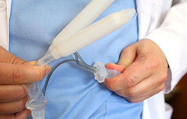
The most common inflatable implant is a 3-piece fluid-filled implant. It is designed to produce a natural-feeling erection and allows a flaccid and "normal" appearance when not having sex. The 3 pieces are: a pair of inflatable cylinders ("tubes") implanted in the penis, a pump in the scrotum, and a water reservoir placed in the lower abdomen. When you squeeze the pump several times, the water from the reservoir is transferred to the cylinders in the penis. The cylinders in the penis are like the inner tubes of a bicycle tire and can be pumped until very rigid. The implant, however, cannot be overinflated and burst. After you and your partner have completed intercourse, you can deflate the device by pressing the deactivation button on the pump.

A malleable penile implant involves a pair of fixed rods that are placed in the penis. The penis is tilted down to urinate and up to have sex. It does not inflate or deflate and remains rigid all the time. For this reason, malleable implants are usually offered only to people who have poor manual function and cannot squeeze a scrotal pump. People who typically benefit from a malleable implant have a spinal cord injury, advanced Parkinson's disease, or advanced rheumatoid arthritis.

A penile implant offers a long-term solution for penetrative sexual relations. The mechanical lifespan of an implant is 10 to 15 years. Once the implant is activated, you can maintain an erection for as long as you desire. You can have spontaneous sex whenever you want and maintain ejaculation and orgasm after the implant surgery. The implant is designed to feel "natural" during sexual intercourse, both for you and your partner.

Any invasive surgery carries risks of infection, bleeding, and surgical mishaps. Infection is very rare today because implants are impregnated with antibiotics, and surgical techniques have improved dramatically over the years.

Retroprostatic and Vesicorectal Urinary Fistulas

A fistula is an abnormal connection between two organ systems. A prostate-rectal fistula is an abnormal connection (hole) between the prostate and the rectum that allows urine to pass freely into the rectum and feces to pass freely into the prostate.

Prostate-rectal fistulas usually occur as side effects of prostate cancer therapy.

Prostate surgery: After or during prostate cancer surgery (open or robot-assisted laparoscopy), a hole may be accidentally created when the prostate is surgically dissected from the rectum. This is a surgical mishap. If the rectal injury is recognized during surgery, it can often be corrected at the time. However, when the injury is not initially recognized and is only recognized days or weeks later, it requires complex surgical repair.

Prostate radiation: Injuries to the rectum and prostate often occur after radioactive seed implants for prostate cancer. Proper placement of the seeds can be complicated. The seeds may be placed too close to the outer edge of the prostate and rectum, and thus the radiation "burns" the tissue, resulting in an ulcer in the prostate and rectum. Over time, the ulcer may open a hole connecting the two organs. Radiation damage continues to worsen over time. A rectal ulcer can be very painful due to chronic infection and can make sitting difficult.

Small fistulas that are only the result of a surgical mishap and have no associated radiation or cryotherapy can often be repaired by a York Mason repair—a rectal division repair of the fistula where an incision in the skin is made in the gluteal fold and deep into the rectal wall.

Another option is to dilate the anus and, through the anus, surgically create a sliding flap of the rectal wall to cover the hole (fistula).

If the fistula is large or there is associated radiation, the fistula repair is very difficult and complicated. Here, the repair usually requires a muscle flap from the leg, transferred to the prostate to act as a patch. If the hole is large, a piece of the lining from the inside of the cheek is also harvested and sewn into the prostate.

Radiation fistulas are the most difficult to repair. The process from start to finish usually requires more than one surgery and a period of at least 6 months.

Treatment usually requires fecal and urinary diversion—a colostomy or loop ileostomy to bring the stool to the skin. This requires the use of a stoma or ostomy bag (appliance) to collect the stool.

Repair of the fistula (hole) requires a muscle flap from the leg or buttock and often a graft from the mouth. After surgery, it takes one or two months for all the tubes to be removed.

Once the fistula is healed, a 3-month or longer interval is allowed before the ostomy (stool bag) is reversed.

Vesicovaginal Fistulas (VVF)

A vesicovaginal fistula is a hole between the bladder and the vagina. Depending on the size of the hole, urine will continuously drain out of the vagina, requiring multiple pull-ups or pads.

In the US, the most common cause of VVF is bladder injury that occurs during a hysterectomy (removal of the uterus). Bladder injuries occur most commonly during open abdominal hysterectomies, followed by laparoscopically assisted vaginal hysterectomies. Bladder injuries during a vaginal hysterectomy are very uncommon.

Most VVFs appear a few days after the hysterectomy. Initially, the leakage from the vagina may be confused with peritoneal fluid leaking from the vaginal cuff.

The diagnosis of VVF is usually made in the office by performing what is called a "tampon test." Here, a gauze pad is placed in the vagina. The bladder is then filled via a catheter with blue-dyed water. A blue-stained vaginal gauze confirms a VVF. In addition to the tampon test, the urologist examines the bladder and vagina with a telescope (cystoscope) and performs a pelvic exam. About 10% of the time, in addition to the VVF, a ureterovaginal fistula—a hole between the ureter and the bladder—also occurs.

How is a ureterovaginal fistula (UVF) diagnosed?
A UVF is usually diagnosed by a CT urogram of the pelvis. If the ureter is not well visualized on the CT, dye may be injected into the ureter via a telescope placed in the bladder. A UVF may also be suggested by the vaginal "tampon test" used to diagnose a VVF—here, the patient is given an oral medication that turns the urine red-orange. If the vaginal gauze turns orange-red, then a UVF is usually also present.

Evaluation of the ureter is always part of the VVF evaluation.

Once the inflammation has subsided after the hysterectomy surgery, a surgical repair of VVF is safe to perform. If the fistula is still inflamed and friable, the success rate of the repair decreases. For this reason, it is important to wait a few weeks before performing the repair. Waiting months for surgery is unnecessary and does not improve success.

There are two main methods of VVF repair through surgery—one through the vagina and the other through an abdominal incision.

Most VVFs can be repaired through a vaginal incision. If the hole is large, a fat pad from the side of the vagina may be harvested and sewn as a patch over the fistula. This fat is called a "Martius flap," and its harvest requires a separate incision on the side of the vagina. If the fistula is also the result of pelvic radiation, a muscle from the leg may be rotated out of the leg and into the vagina to act as a patch.

Very high fistulas in the vagina and/or near the ureter in the bladder usually require an abdominal approach because the hole may be difficult to reach through the vagina. If the ureter is involved, it may need to be sewn back into the bladder—which is much easier to do through the abdomen. If the VVF is repaired through an abdominal incision, the fistula is closed with sutures. The fistula is also patched with a fat pad taken from the stomach.

A UVF is usually repaired by cutting the ureter and sewing it back into the bladder. In selected cases, a stent (plastic tube) may be placed through the bladder to the cut end of the ureter and into the kidney. If this stent can be placed, the UVF usually heals without the need for major surgery.

Urethral Stricture (Urethral Narrowing)

The urethra in men is the tube that carries urine from the bladder to the outside of the body and also serves as the channel through which semen is ejaculated.

The anterior urethra is the portion of the urethra that runs from the tip of the penis to just before the prostate. The posterior urethra is the part of the urethra that passes through the prostate and the external sphincter valve.

The relative location of the urethra within the spongy tissue (erectile tissue surrounding the urethra) changes along the divisions of the urethra. The anatomical location of the lumen (urethral cavity) in relation to the spongy tissue is crucial for selecting the type of surgical correction and determining the internal surgical incisions through a cystoscope (telescope to look inside the urethra).

An anterior urethral stricture is a scar of the inner lining of the urethra that also commonly extends into the surrounding erectile tissue (corpus spongiosum).

The scar tissue (stricture) can contract in all directions, shortening the length of the urethra and narrowing the diameter of the urethra. As the urethra narrows, the urine stream slows down.

Most current urethral strictures are the result of blunt trauma to the perineum (the area between the thighs from the end of the spine to the pubic bone), such as a straddle injury, or previous urethral instrumentation by a doctor or nurse, or a chronic indwelling Foley catheter.

Inflammatory strictures, such as those secondary to venereal diseases like gonorrhea or chlamydia, are very rare today. In the US, the most common cause of inflammatory strictures is lichen sclerosus and atrophicus (commonly known as LSA), where whitish plaques affect the glans (head of the penis), meatus (urinary opening), and foreskin. It is also a common cause of phimosis (infection of the foreskin) and is therefore often seen at the time of adult circumcision. LSA begins as inflammation of the head of the penis that severely narrows the urethral opening at the tip. High-pressure urination and infected urine accumulate behind the narrowing, and then the narrowing descends down the urethra. Over time and neglect, a very long urethral stricture can occur. These strictures are difficult to "fix."

As the urethral lumen (tube) gradually narrows, patients begin to suffer from weak urine flow, straining to urinate, urinary spraying, hesitation, incomplete emptying, urinary retention (inability to urinate), and post-urination dribbling. Frequent and painful urination are also common initial complaints.

On palpation, the urethra can often feel firm in the area where the worst aspects of the scar tissue of the corpus spongiosum, which surrounds the urethra, are located. A tender mass along the urethra is usually an abscess (pocket of infection or pus).

An office test that helps diagnose a urethral stricture is called a urinary flow rate. It is a large funnel-shaped machine that, when urinated into, calculates the time of urination and the maximum speed at which urine is exiting the penis. Peak urinary flow rates below 10 milliliters/second indicate a very slow flow and, therefore, significant blockage.


Retrograde urethrography (X-ray of the urethra) and voiding cystourethrography (X-ray of the bladder and urethra during urination) are the gold standard X-rays for evaluating the length and location of the stricture and its functional significance. The X-rays provide the surgeon with a map of the urethra and thus allow them to decide on the best treatment for a potential cure. The X-ray involves placing a small tube at the tip of the penis and injecting X-ray contrast into the urethra. The bladder is then filled with contrast, and the patient is asked to stand and urinate. X-rays are taken of the urethra during urination.

Urethral stricture X-ray

Ultrasound is another method of evaluating the urethra. It is particularly useful for imaging the bulbar urethra (the part of the urethra between the scrotum and the anus). Here, a sterile gel is injected into the urethra, and an ultrasound probe is placed on the skin at the same time. This procedure takes only a few minutes and is performed in the office.

Cystoscopy: A telescope to look inside the urethra is another important part of the evaluation. Cystoscopy is an important complement to urethral X-rays, as it allows a view of the color, actual location, and a more accurate measure of the stricture's rigidity. A pediatric cystoscope is also often used to examine and pass through the stricture. The pediatric scope is very narrow in width and thus very useful for better assessing the length and location of the stricture—especially when other imaging results are ambiguous.

Urethral calibration: Serial metal instruments are often inserted into the urethra to accurately determine the caliber or size of the urethra. This is typically done in the office and takes less than a few minutes to perform.

- Urethral discharge.
- Urinary tract infection.
- Bladder infection and inflammation.
- Prostatitis (inflammation/infection of the prostate).
- Epididymitis (inflammation/infection of the epididymis; a duct system that stores sperm during maturation).
- Abscess in the tissue around the urethra.
- Urethral diverticulum (abnormal opening of a pouch in the urethra)—which can predispose to chronic infection and stone formation.
- Urethrocutaneous fistula (a hole in the urethra that goes to the skin).
- Urethral cancer—one-third of men with urethral cancer have a history of stricture.
- Bladder stones (due to chronic urine accumulation and infection).


The goal of stricture management is cure and not just temporary control. Open surgical urethroplasty (scar excision surgery) has a long-term success rate of approximately 80 to 95 percent and should be considered the gold standard against which all other methods are evaluated.

In general, dilation is only a management tool and not a cure. This is usually reserved for patients who are not candidates for more invasive surgical intervention.

The less traumatic and safer methods are serial catheter dilation over several weeks or balloon dilation.

Dilation potentially cures only web-like strictures, with minimal or no scarring of the surrounding tissue.

Overall, long-term success is low, and recurrence rates are high.

Internal urethrotomy encompasses all methods of surgical incision or ablation to open a stricture that are performed through a telescope placed in the urethra. No skin incision is made here.

At best, the goal of urethrotomy is to create a larger-caliber stricture that does not obstruct urination.

Urethrotomy is potentially curative only for very short strictures (less than 1 cm) that have minimal or no surrounding urethral scar tissue.

For strictures in the bulbar urethra less than 2 cm in length, at one year, the success rates after the first urethrotomy are 60 percent, and at five years, success drops to the range of 26 to 14 percent.

Repeating urethrotomy typically has zero% long-term success.

Laser urethrotomy seems attractive and would improve the mediocre results of knife urethrotomy. However, the results are no better than standard techniques.

The side effects of urethrotomy are potentially: obliteration of the lumen (cavity), as well as hemorrhage (heavy bleeding), sepsis (a serious reaction to infection throughout the body), incontinence (urine loss), and a very rare and transient erectile dysfunction and glans numbness after urethrotomy.

Urethroplasty

In general, if you feel minimal pain and are not taking any painkillers, you can drive. Use common sense.

We recommend taking 3 to 4 weeks off work, especially if you do manual labor or lift heavy objects for a living. In general, it is safe to return to work once the catheter is removed. However, if you have an administrative job, you can likely return to work after 1 to 2 weeks. We suggest that you consider working fewer days in the first week of your return to work.

What is urethroplasty? "Urethro" (urethra) + "plasty" (to repair) = to repair the urethra.

Urethroplasty is the surgical reconstruction, either open/robotic or replacement, of a narrowed and scarred urethra. Urethroplasty is considered the gold standard for urethral reconstruction with the best and most lasting results.

Before any urethroplasty, the scar must be stable and no longer contracting. Thus, it is preferable that the urethra not be dilated or cut for three months before the planned definitive surgery. If the patient with a stricture goes into urinary retention before the surgery date, a suprapubic tube is usually placed. A suprapubic tube, also known as an SP tube, is a small tube that is placed through the skin and directly into the bladder. The tube is typically 2 to 3 cm above the pubic bone.

For short strictures involving the bulbar urethra (the part of the urethra under the scrotum and up to the prostate), a segment of the scarred urethra can be completely removed, and then the two cut ends of the urethra are sewn together.

Excising a small segment of the urethra and sewing the ends together typically has the best long-term surgical results—approaching 95%.

Excision by strangulation cannot be performed on the penile urethra or for long strictures of the bulbar urethra because there will be too much tension in trying to bring the two cut ends together. Tension on the suture line leads to surgical failure and potentially penile shortening and curvature.

A graft is a piece of tissue that is transferred from one part of the body to another. A graft does not have its own blood supply, so it depends on the host's blood supply (where it is transferred) to survive.

Typical grafts used to reconstruct the urethra are harvested from extra-genital skin or the inner lining of the mouth. The mouth graft is known as a "buccal graft."

Grafts are used to reconstruct the narrowed urethra by enlarging the size of the urethra, patching it, rather than a total replacement.

Grafts are highly effective in the bulbar urethra as an onlay or patch technique. The cheek lining is typically used as a patch graft because it is easy and quick to harvest, has a hidden suture line, does not contract much, and can result in durable success rates approaching 80-85%.

Skin grafts used in urethral reconstruction can shrink up to 50 percent and thus result in lower success rates than the buccal (oral) graft. Penile skin should be avoided as a graft when the penile skin is not very redundant or elastic, or when the penis is affected by Lichen Sclerosus (a skin disease).

A flap is a transfer of tissue from one part of the body to another, where the donor's blood supply remains intact. In other words, a penile skin flap used to reconstruct a urethral stricture does not depend on the scarred urethra for its survival. The penile skin flap is good for reconstructing long strictures of the penile urethra. Penile skin flaps are versatile and are used as a patch for the narrowed segments of the urethra. Short-term success rates approach 80-85%. Flaps that are rolled into a tube have a failure rate of nearly 50%—therefore, flaps are reserved for patches and not for replacement.

Scrotal skin flaps should be avoided in urethroplasty, as their complication rate is high, and their success rates are low.

For patients who have failed previous urethroplasties or where the urethra and local skin are severely scarred, staged urethroplasty is usually indicated. Here, the scarred urethra is typically surgically excised, and in its place, a buccal or skin graft is placed. This replaced urethra is left open to the air to heal over the next few months. As the meatus (urine hole) is usually placed in front of or under the scrotum, these patients need to sit to urinate for a few months. Once the graft is soft and well-healed, the patient undergoes a second surgery ("staged") to roll the graft into a tube to reconstruct the urethra. A staged urethroplasty can often have more than two stages and require more than one graft phase. Staged urethroplasty is usually reserved for the worst urethral strictures.

One option is not to reconstruct the urethra and instead bypass the urine. Here, the urethra is opened under the scrotum, and a skin flap is sewn into the urethra. Thus, with the bulbar urethra sewn to the skin, urine can easily exit. However, the patient will need to sit to urinate. This is a reasonable solution for a severely scarred urethra in an older patient.

The other option is to use a combination of a penile skin flap for the penile urethral stricture and a buccal graft for the bulbar urethra under the scrotum to reconstruct the narrowed urethra.

Urethral injuries that occur from a pelvic fracture are not really strictures but rather distraction injuries—where the urethra is torn at the junction of the prostate and the membranous urethra or at the junction of the bulbar urethra and the membranous urethra. Typically, the space between the torn ends of the urethra is approximately 2 cm.

Typically, the urethra is not repaired at the time of the pelvic fracture—since there are more life-threatening injuries. To divert urine, a tube is usually placed through the lower abdominal skin and directly into the bladder (known as a suprapubic tube). After the patient heals from the pelvic fracture, the urethra is reconstructed. This usually occurs 3 or more months after the accident.

Although newer imaging methods, including ultrasound and magnetic resonance imaging (MRI), are sometimes used for evaluation, cystography (X-ray of the bladder) and retrograde urethrography (dye injected into the penile urethra) remain the gold standard for evaluating posterior urethral strictures.

In addition to the radiographic study, visualizing the stricture from above and below is important. This is done with an office cystoscopy (telescope) through the penile urethra and through the bladder.

Open one-stage urethroplasty, where the scarred segment is excised, and the two ends of the urethra are sewn together, is the gold standard for posterior urethral stricture repair. Long-term success rates approach 90 to 95 percent.

Grafts are rarely used due to insufficient blood supply.

The results of minimally invasive surgical techniques (via a telescope) are generally very poor and should be considered only a temporary measure and never a cure.

Once the scarred tissue is removed, the two ends of the urethra can be brought together based on the natural elasticity of the urethra. By releasing the urethra from its underlying attachments, the urethra is very elastic and can stretch up to 70% of its length.

Occasionally, the distance between the ends of the urethra is too great—and the elastic urethra will not bridge the gap. In these cases, a small piece of the pubic bone is removed. Removing part of the pubic bone gives the urethra a direct shot to the prostate and thus shortens the distance.

Before any urethroplasty, the scar must be stable and no longer contracting. Thus, it is preferable that the urethra not be dilated or cut for three months before the planned definitive surgery. If the patient with a stricture goes into urinary retention before the surgery date, a suprapubic tube is usually placed. A suprapubic tube, also known as an SP tube, is a small tube that is placed through the skin and directly into the bladder. The tube is typically 2 to 3 cm above the pubic bone.

As with most surgeries, the pain is worst in the first 12 hours and then gradually decreases. By the morning after surgery, you should feel quite comfortable. The pain is usually just a constant ache that lasts a few days.

If you experience sudden pain in the penis, pubic bone, or lower abdomen, this is "bladder spasm" or catheter pain. This is not the pain from your surgery. To treat "bladder spasm" pain, you can take medications that calm the bladder, such as Ditropan, Levsin, or Detrol.

The surgery time varies because each surgery and patient are unique. In general, the procedure takes three hours but can range from 2 to 5 hours, depending on the severity of the scar tissue and the extent of the reconstruction.

Almost all patients are discharged immediately after surgery or within 23 hours. A small percentage of patients may stay an extra day if they have no help at home or if they need to travel a long distance by car.

Ureteral Strictures

The ureter is the duct that carries urine from the kidney, where it is produced, to the bladder, where it is stored until urination. Ureteral strictures or narrowings can be primary (congenital or present at birth, such as Ureteropelvic Junction Stenosis/UPJ) or acquired over time due to trauma, endourological surgeries for kidney stones, neoplasms, among others.

Rarely are primary causes diagnosed in adulthood, as they are typically managed surgically in childhood through pyeloplasty. Secondary causes are usually more complex because they are associated with injuries, infections, and scarring. Surgical correction varies from grafts using advanced robotic surgery to replacing the ureter with segments of the gastrointestinal tract.

Professor Tiago Rosito is a pioneer in Brazil in the use of minimally invasive methods for managing ureteral strictures, whether through conventional laparoscopy or robotic technology.